Bioimaging Services for Nanoparticles
With the development of nanomedicine, the application of nanomaterials to tumor-targeted drug therapy is expected to improve the effectiveness of chemotherapy. Imaging and quantitative detection of nanoparticles in tissue cells can help to further localize and quantify drug concentration and distribution in tissue cells, providing visualization of drug effects. These imaging techniques enable scientists to study drug release kinetics, release mechanisms, targeting efficiency, and therapeutic effects, helping researchers gain a deeper understanding of biological pathways, disease progression and the impact of experimental drugs. It's critical for translational research and early clinical stage validation of drug bioavailability, permeability, target accumulation and mechanism of action. Currently, the representative imaging modes in preclinical or clinical practice are magnetic resonance imaging (MRI), optical fluorescence imaging and photoacoustic (PA) imaging, computed tomography (CT), positron emission tomography (PET).
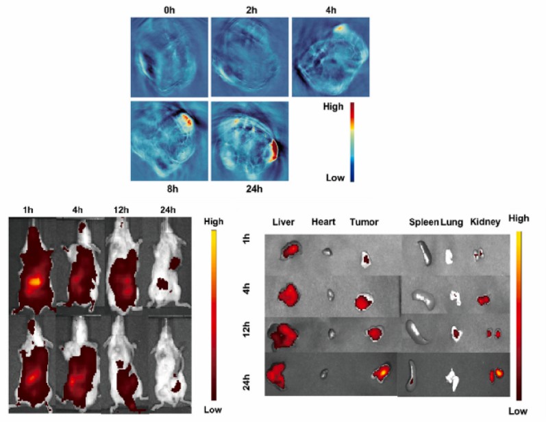 Figure 1. Visualization of imaging maps inside and outside the organism.1
Magnetic Resonance Imaging (MRI)
Magnetic nanoparticles are nanoscale particles with magnetic properties that can be imaged in response to an external magnetic field. A key advantage of MRI is that the signal is not limited by tissue scattering, allowing imaging of areas deep within the body. Common magnetic imaging materials include ferric oxide (Fe3O4), small molecule gadolinium(III) chelates, and manganese-based nanomaterials. Most notably, superparamagnetic iron oxide (SPIO) nanoparticles have been shown to exhibit a reduction in the T2 signal of tissues and have received FDA approval for use in humans in some clinical applications.
Experimental Method
1. Preparation of samples containing magnetic properties
2. Animal model drug administration
Use ether to anesthetize the experimental animals, and administer the drug by intraperitoneal injection or other means.
3. Instrumental imaging
Use MRI equipment to perform dynamic scanning of the experimental animals with and without drug administration.
4. Graph processing and analysis.
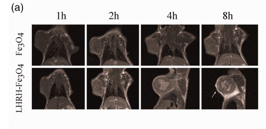 Figure 2. The imaging pictures of Fe3O4 in breast cancer.2
Photoacoustic Imaging (PAI)
Some metallic nanomaterials and carbon-based nanomaterials can be capable of converting light energy into heat energy under near-infrared light (700-1000 nm) irradiation to produce a strong PA signal. Common luminescent nanomaterials include gold nanorods, metal sulfides, melanin and upconversion nanoparticles.
Experimental Method
1. Preparation of nanomaterials for optical imaging.
2. In vitro imaging
The cell culture was affixed to the wall and co-incubated with the sample for a certain time, and after the cells ingested the sample, the cells were gently washed to remove the ungrafted sample. The imaging pictures were examined using a multispectral photoacoustic tomography imaging system (MSOT).
3. In vivo imaging
In vivo PA imaging, the animals were anesthetized by an isoflurane-air mixture (2% isoflurane by volume) and placed in a horizontal position prevented by a thin PE membrane from contacting water. Then 100 µL of nanoparticles (at a certain concentration) were injected into the experimental animals. A series of cross section images were acquired along the axial direction using MSOT imaging system.
 Figure 3. Photoacoustic imaging of gold nanorods in vivo.3
Fluorescence Labeling Imaging (FI)
Fluorescent substances are compounds with the chemical structure of a co-rotating double bond system, which can be excited into an excited state when irradiated by ultraviolet light or blue-violet light, etc., and emit fluorescence when recovering from the excited state to the ground state. Fluorescent labeling technology refers to the use of fluorescent substances covalently bound or physically adsorbed to a group of the molecule to be studied. The fluorescent properties are used to provide information about the object under study. Common fluorescent dyes include fluorescein isothiocyanate (FITC) dyes, NIR dyes (AIE photosensitive molecules), DIO, CY3, CY5, TRITC, luciferase, indocyanine green, methylene blue, coumarin and others.
Common fluorescent dyes luminous colors:
| Name |
Lighting Color |
| FITC, ICG Series, DIO |
Yellowish green to green |
| CY3, TRITC |
Dark orange |
| CY5, luciferase, Coumarin, Methylene blue |
Blue to blue-purple |
Experimental Method
1. Raw material preparation: labeling nanomaterials with fluorescent dyes.
2. In vitro imaging
The cells were cultured and affixed to the wall and then co-incubated with the sample for a certain time. After the cells ingested the sample, the cells were gently washed to remove the ungrafted sample. The excitation and emission wavelengths of the fluorescent dyes are set, and the fluorescence of the cells is observed by confocal laser scanning microscopy (CLSM).
3. In vivo imaging
For in vivo imaging, inject 100µL of fluorescent dye-NPs solution (concentration to be determined) into the experimental animals. Set the corresponding excitation and emission wavelengths, and detect the distribution of NPs in vivo with the IVIS imaging system.
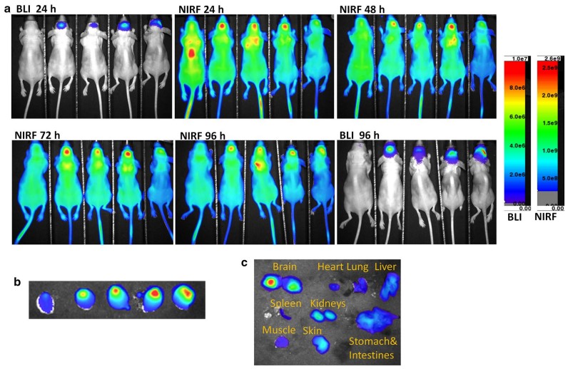 Figure 4. Fluorescence imaging of phospholipid nanoparticles labeled with IR780 dye.4
PET and CT Imaging
PET is generally radioisotope labeled imaging, and common radioisotopes include 18F, 64Cu, and 76Br. CT is often used with PET, which uses a series of X-ray images to create a three-dimensional image from multiple perspectives to reveal details about the anatomical features of the tissue of interest.
Experimental Method
1. Raw material preparation: labeling nanomaterials with radioisotopes.
2. Imaging
The experimental animals were injected with isotope-labeled nanomaterials and imaged using a microPET/microCT Inveon rodent model scanner.
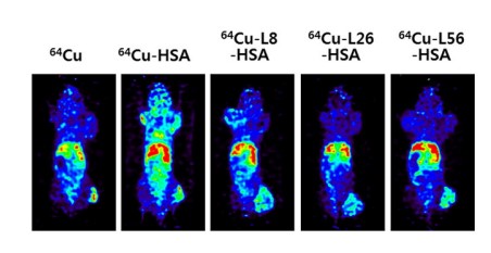 Figure 5. Fluorescence imaging of albumin nanoparticles labeled with 64Cu in vivo in mice.5
Quantitative Detection of Nanoparticle
Imaging as well as quantitative detection of drug molecules in vivo can help researchers to understand drug release kinetics and therapeutic efficacy and further optimize drug dosage administration. For small molecule drugs and achieving quantitative detection on tissue sections, LC-MS/MS/LC-HRMS/ICP-MS is usually utilized.
Conclusion
The qualitative and quantitative study of nanoparticles in vivo at the tissue, cellular and molecular levels is based on the principle that light can penetrate the tissues of experimental animals and that the intensity of light can be quantitatively detected by the instrument. The various instrumental methods have their own advantages and disadvantages, and CD Bioparticles can provide you with a more expert judgment based on your material.
| Methods |
Advantages |
Disadvantages |
| MRI |
Without utilizing any radiation, it is possible to reconstruct three-dimensional images of the entire organ and analyze functional parameters. |
The sensitivity of MRI is relatively poor. Even with the use of strong contrast agents, the sensitivity of MRI is significantly lower than PET and fluorescence microscopy. |
| PAI |
Possesses good temperature sensitivity, high imaging accuracy and speed for real-time monitoring. |
The light is scattered and absorbed strongly in the tissue, and the penetration ability is poor. |
| FI |
Lower cost, higher sensitivity, can do multicolor imaging. In addition NIR dyes can do deeper tissue imaging |
Some dyes suffer from photobleaching. Some fluorescent dyes have some toxicity and poor tissue penetration. |
| PET and CT |
Good penetration and good imaging of hard tissue. |
Strong ionizing radiation. |
Quotations and Ordering

References
- Yu C, et al.; Photoacoustic imaging-guided triple-responsive nanoparticles with tumor hypoxia relief for improving chemotherapy/ photothermal/photodynamic synergistic therapy against breast cancer. Biomedicine & Pharmacotherapy. 2023, 164: 114928-114942.
- Dian D, et al.; Application of luteinizing hormone-releasing hormone-ferrosoferric oxide nanoparticles in targeted imaging of breast tumors. Journal of International Medical Research. 2019, 47:1749.
- Shrestha B, et al.; Gold nanorods enable noninvasive longitudinal monitoring of hydrogels in vivo with photoacoustic tomography. Acta Biomaterialia. 2020, 117:374.
- Li S, et al.; Near infrared fluorescent imaging of brain tumor with IR780 dye incorporated phospholipid nanoparticles. Journal of Translational Medicine. 2017, 15(18): 6919.
- Kang K, et al.; Albumin as natural nanoparticles for in vivo imaging and drug delivery. Nanomedicine: Nanotechnology, Biology and Medicine. 2018, 14(5): 1782.
