Bioparticle Component Analysis
CD Bioparticles offers custom services to characterize bioparticles using advanced microscopic techniques. Our experienced scientists and technicians have created a comprehensive platform to measure different bioparticle components.
Bioparticle Component Analysis Introduction
Bioparticle component has been found to have a pround effect on stability and the feature of bioparticle, therefore it becomes crucial to measure bioparticle component to make sure bioparticles are in accordance with the formulation which is designed to meet drug delivery requirement. The efficiency of drug delivery is determined by the transport carrier and the drug components. The carrier component further determines the character of the drug delivery system and the toxicity assessment of the transport system. For example, in a liposome transport system transport system, liposome component is an important parameter for subsequent drug embedding and delivery. Different lipid components are selected to form different liposomes depending on the drug delivery requirements and the nature of the drug. In addition, long-term storage of biological particles requires the detection of their constituents before use to ensure that the components are not oxidized or degraded.
Bioparticles Component Analysis Methods
X-ray Diffraction (XRD)
XRD is the most common method utilized for determining the atomic and molecular structure of materials. The crystalline atoms lead to the diffraction of the incident X-ray beam into specific directions. Therefore, the diffraction angles and corresponding intensities can be measured and recorded, which depict a three-dimensional picture of the density of electrons within the crystal. A great number of crystalline materials can be characterized by XRD, including inorganic, organic materials and biological molecules. The physical states of materials for XRD measurement are flexible, they can be loose powders, thin films, polycrystalline and bulk materials. Our scientist team and advanced facilities allow us to provide XRD measurements with advantages including minimum quantity of sample required, non-destructive measurement, and easy to interpret.
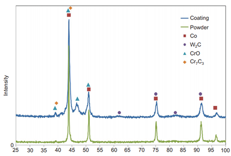
Figure 1. X-ray diffraction (XRD) analysis. (Rocco L, et al. Light: Science & Applications. 2012, doi:10.1038/lsa.2012.10)
Liquid Chromatography-mass Spectrometry (LC-MS/MS)
Organic component analysis and inorganic elemental analysis can be evaluated by using liquid chromatography-mass spectrometry (LC-MS). It can provide information about the identification, quantitation, and mass analysis of the materials and compounds. LC-MS first separates compounds in sample mixture via the chromatography based on the compound intrinsic affinity for the stationary phase and mobile phase. When the separated compounds pass through the mass detector, the intensity is collected and calculated for each compound as it is vaporized and atomized in the plasma by comparing to a standard reference. The mass fraction or size of the compounds can be correlated to the measured data point. Our LC-MS/MS service has the capability to provide information about the identification, quantitation, and mass analysis of each component. Data analysis service is also provided upon request.

Figure 2. Positive ion electrospray tandem mass spectrometry (Cong W, et al. Journal of Biomolecular Screening. DOI: 10.1177/1087057113492852)
Raman Spectroscopy
Raman spectroscopy is commonly used in chemistry to provide a structural fingerprint by identifying molecules. It relies on inelastic scattering (or Raman scattering) of monochromatic light usually from a laser in the visible, near infrared, or near ultraviolet range. The laser light interacts with molecular vibrations, phonons or other excitations in the system, resulting in the energy of the laser photons being shifted up or down. The shift in energy gives information about the vibrational modes in the system. Infrared spectroscopy yields similar, but complementary, information. Typically, a sample is illuminated with a laser beam, then electromagnetic radiation from the illuminated spot is collected with a lens and sent through a monochromator. Elastic scattered radiation at the wavelength corresponding to the laser line (Rayleigh scattering) is filtered out by either a notch filter, edge pass filter, or a band pass filter, while the rest of the collected light is dispersed onto a detector. Our Raman spectroscopy service has the capability to collect the information of structural fingerprint used to identify bioparticle components.
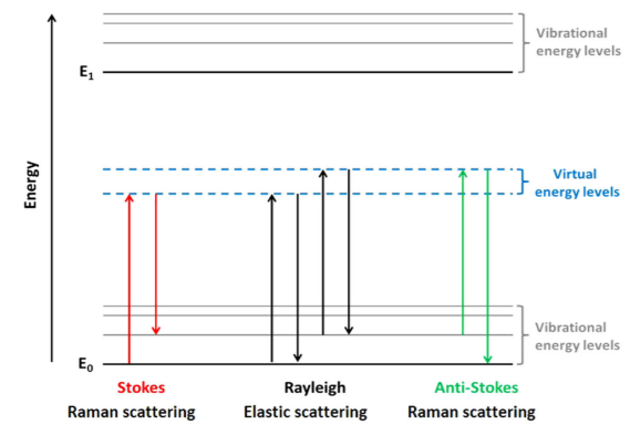
Figure 3. The date of Raman scattering. ( KJI Ember et al. Raman spectroscopy and regenerative medicine. 2017, doi:10.1038/s41536-017-0014-3)
CD Bioparticles offers a full set of service for bioparticle component analysis when drugs are encapsulated into bioparticles. For more detailed information, please feel free to contact us or directly send us an inquiry.
Quotations and Ordering

References:
1. Gardiner, D. J., Graves, P. R. Practical Raman spectroscopy. Springer-Verlag. 1989, ISBN 978-0-387-50254-0.
2. Pitt, J. J. Principles and Applications of Liquid Chromatography-Mass Spectrometry in Clinical Biochemistry. The Clinical Biochemist Reviews. 2017, 30 (1): 19–34.
3. Compton A. A Quantum Theory of the Scattering of X-rays by Light Elements. 1923, Phys. Rev. 21 (5): 483.
