Bioparticle Size and Morphology Analysis
CD Bioparticles offers custom services to characterize bioparticles using advanced microscopic techniques. Our experienced scientists have created a comprehensive platform to measure different bioparticles size distribution, in the meantime also analyze their morphology.
The Importance of Bioparticles Size in Drug Delivery System
Bioparticle size has been found to have a profound effect on drug release, therefore it becomes crucial to measure particle size distribution after formulation to make sure bioparticles meets drug delivery purpose. Bioparticle morphology, another important parameter for bioparticles characterization, could also be visualized when electron microscopy applies. Theoretically, smaller particles lead to fast drug release since it offers a larger surface area to expose drugs to the system. On the contrary, larger particles come with slow drug diffusion. However, smaller bioparticles tend to aggregate during storage and transportation. Therefore, a mutual balance between small size and maximum stability of nanoparticles should be reached. Different types of bioparticles might have different size characters, and also size monitoring before and after drug encapsulation is a prerequisite step of quality control for a successful formation development in drug delivery.
Bioparticles Size Analysis Methods
Dynamic Light Scattering (DLS)
Dynamic light scattering (DLS) is the fastest and most popular technique for accurate estimation of the particle size and size distribution. A solution of spherical particles in Brownian motion causes the changing the wavelength of the incoming light when the light hits the moving particle. Because this change is related to the size of the particle in solution, it has been used to extract the information of size distribution. Our robust DLS platform could provide accurate, reliable and repeatable particle size measurement in one or two minutes. Our advanced technique could allow size measurement from 10um to < 100nm with a low sample volume requirement.
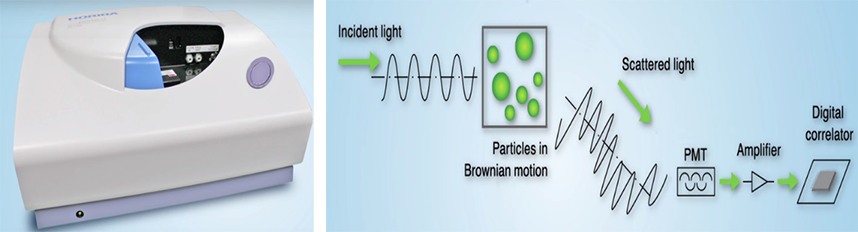
Figure 1. Overview of dynamic light scattering mechanism.
Scanning Electron Microscopy (SEM)
Scanning Electron Microscopy could be used to determine the size, shape and surface morphology of nanoparticles with direct visualization of a sample by scanning the surface with a focused beam of electrons. Sample preparation for SEM involves sample drying, mounting, and coating. The surface characteristics of the sample are obtained from the emitted electrons from the sample surface after scanning with a focused beam of electrons. An average mean size value could be evaluated by SEM, but limited information about the size distribution and true population average is given by SEM. In addition, these techniques are time-consuming, costly due to the sample preparation process and electron beams. We offer comprehensive services to help you from sample preparation to image analysis. Our scientist team and advanced facilities allow for samples observing in high vacuum, in low vacuum or wet conditions in variable pressure together with a wide range of temperatures with specialized instruments.
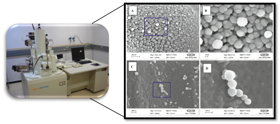
Figure 2. Scanning electron microscopy (SEM) instrument and images of polymeric nanoparticles. (Alexander Vakurov, et al. J Nanopart Res (2012) 14:1302)
Transmission Electron Microscope (TEM)
TEM also provide sample visualization with a different principle compared to SEM by obtaining a transmitted beam of electrons through samples. Ultra-thin samples preparation required for electron transmittance is complicated and time-consuming. The nanoparticles dispersion is fixed by either a negative staining plastic embedding. Besides, cryo-EM has become a revolutionary method to visualize nanoparticles and size distribution analysis because it allows the observation of samples that have not been stained or fixed, maintained in their native environment instead. Cryo-EM is very powerful but also with a very high cost. Our high energy electrons (80keV-200keV) TEM platform could help our client to collect high resolutions images around 1-2Å, provide tailored service to characterize different types of nanoparticles.
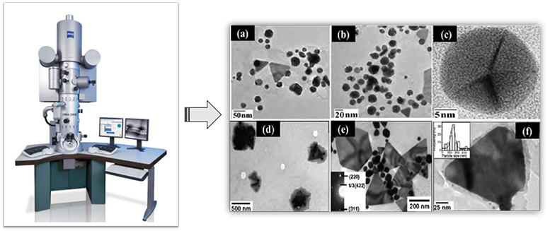
Figure 3. Transmission electron microscopy (TEM) image of nanoparticles (A. Rai, et al., Mater. Res. Bull. 42, 1212 (2007)).
Atomic Force Microscopy (AFM)
Atomic force microscopy (AFM) offers accurate measurement of size and size distribution by tapping the probe on to the surface across the sample. A topographical map of the sample is obtained based on forces between the tip and the sample surface. Without any specific treatment, AFM is able to image delicate biological and polymeric nano and microstructures without disturbing the samples. Our AFM service has the capability for high resolution topographical imaging of samples in air or liquid environments. Also, real-time changes in sample morphology or structure could be monitored by time-lapse experiments. Data analysis service is also provided upon request.
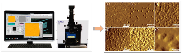
Figure 4. Atomic force microscopy images showing the formation and size changes of Cp–Ag nanoparticles. (Subhadip Ghosh, et al., Analyst, Issue 15, 2013.)
Quotations and Ordering

References:
1. Rai, A., et al. Synthesis of triangular Au core-Ag shell nanoparticles. Mater. Res. Bull. (2007) (2007) 42, 1212.
2. Linda E. F., et al. Transmission electron microscopy as a tool for the characterization of soft materials: application and interpretation. Adv. Sci. (2017) 4, 1600476.
